Properties of Yeast Prions
One of the most well-studied prions is called [PSI+] (pronounced like “sigh”). The [PSI+] prion occurs as a result of an altered conformation of the Sup35 protein. Sup35 is one of the two known yeast translation termination factors. When Sup35 is soluble in the [psi-] state, it mediates efficient translation termination at all 3 stop (nonsense) codons in yeast (UAA, UAG, UGA).
In the [PSI+] state, however, Sup35 is aggregated and unavailable for function – resulting in read-through of nonsense codons and producing elongated polypeptide chains. We can follow the [PSI+] phenotype in yeast by using an adenine marker (ade1-14 mutation) that harbors a premature nonsense codon. When translation termination is efficient, the ADE1 gene product is not generated, the cells are phenotypically ade-, and the cells become red due to the accumulation of a metabolic intermediate in this pathway. However, when the cells are [PSI+], the ADE1 gene product is translated completely due to the read-through of the nonsense codon. Therefore, the [PSI+] cells are phenotypically ADE+ and they remain white. Since the prion state is self-replicating, this results in dominant, cytoplasmic inheritance. As such, we refer to prions in yeast as epigenetic elements of heritability.
One of the advantages to studying prion propagation in yeast is the ability to perform genetic screens to identify modifiers of this propagation. A genetic screen revealed that a chaperone protein called Hsp104 can influence prion propagation. We can delete Hsp104 in yeast and prion propagation is disrupted.
We can also observe the protein conformational transitions that correspond to the PRION+ state of a protein in yeast by making use of a GFP fusion protein. When this fusion of the prion protein to GFP is expressed in cells that are [PRION+], we see the aggregation pattern because the GFP fluorescence is in tight foci. However, when we express this fusion protein in [prion-] cells, the pattern of fluorescence remains diffuse.
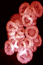
Additionally, we can biochemically show an altered conformational state of prion proteins as well. In one assay, we subject the cell lysates to a high speed centrifugation step and then the supernatant and pellet fractions are analyzed by western blot. In the prion- state, the protein is largely in the supernatant fraction. In the PRION+ state, however, the prion protein is mostly in the pellet. Biochemical research has demonstrated that many yeast prion proteins readily form amyloid-like fibers as shown in the EM photograph at the top of the page. These fibers are reminiscent of those observed with several misfolded proteins that are associated with a number of neurodegenerative disorders in humans. This suggests that yeast prion proteins are a tractable model system to study the same type of protein aggregation that occurs in human diseases.
 For information on projects currently underway, go to the
Members page.
For information on projects currently underway, go to the
Members page.